Fabrice Ducrest © UNIL
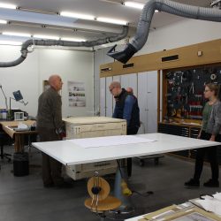
Since the West Swiss unit of the Swiss Institute for Art Research (SIK|ISEA) at UNIL has no laboratory, the team in charge of the globes asked the restorers at the institute’s main site in Zurich to take over. In autumn 2014 Margaux Genton, an assistant restorer, came to Lausanne for a preliminary examination of the globes in situ. Having already restored a 17th-century celestial globe, she had both the experience and the knowledge needed for this type of job.
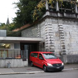
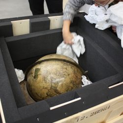
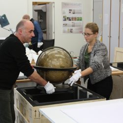
It was agreed that the globes would have to be taken to the SIK|ISEA’s laboratories in Zurich for additional analyses (including a C14 test of the celestial globe using a new, non-invasive method) and then be cleaned and consolidated with a view to conserving and exhibiting them. The transfer took place on 10 November 2014.
-

-
© Micheline Cosinschi, UNIL
-

-
© Micheline Cosinschi, UNIL
-

-
© Micheline Cosinschi, UNIL
-

-
© Micheline Cosinschi, UNIL
SIK|ISEA was asked to perform the following tasks :
- assess the various analyses conducted hitherto,
- and summarise the results,
- find out more about the globes’ constituent materials and construction using various other methods of observation and analysis,
- assess the globes’ state of conservation,
- consolidate and restore them.
To accomplish this mission, SIK|ISEA used new methods of observation and analysis which are listed below.
| Method |
Parameter |
| Visual observations |
Visible light and grazing incidence light |
| Binocular magnifier |
Zeiss technoscope, enlargement 4x-105x |
| Ultraviolet fluorescence (UVF) |
Source of rays : (black light 320-400 nm) Dr Hönle, UVAHAND 250 / UVASPOT 400T
Camera : Nikon D 600 modified for 350-1100 Nm spectral range
Filters : IR NG Makario (Bandpass 400-700 nm) neutralisation filter + Kodak Wratten E2 (Longpass 420 nm)
|
| Infrared reflectography (IR) |
Source of rays : variable-intensity halogen lamps
Camera : Nikon D 600 modified for 350-1100 nm spectral range
Filter : Longpass 700 nm / 830 nm
|
| X-ray with X-rays |
Machine : Gilardoni (Art-Gil, 5 mA)
Films : Agfa Strukturix D4 DW
|
| Fournier-Transformed Infrared Spectroscopy (FTIR) |
Perkin Elmer System 2000 with IR/vis (Perkin Elmer i-series) microscope
CVD diamond-coated steel cylinder
|
| Raman spectroscopy |
Renishaw inVia Raman Microscope (01/2007)
Laser 785 nm (Diode) : Renishaw HP NIR785 (300 mW)
Laser 633 nm (Gaz) : Renishaw HeNe 633 (17 mW)
Laser 514 nm (Gaz) : Spectra-Physics Ar ion laser (24 mW) |
| Direct temperature mass spectrometry (DT-MS) |
DSQ II-Thermo Electron Instrument
Heating speed : 10° C/s (up to 1000° C)
EI 16 eV
Quadrupole Mass Spectrometer
Area of measurement : 45-1050 m/z
|
| Stratigraphic cross-sections |
Resin : CEM 4000 Lightfix, solidification in blue light
Dry slicing
Micromesh polishing (up to 12000 = P1400 = 2-6 ?m granularity)
Microscope Zeiss AXIO Scope A1, different lighting modes
|
| Variable-pressure scanning electron microscope (VP-SEM) |
Zeiss energy dispersive analysis system EVO MA 10 (2014)
High vacuum mode 10e-5 Pa, low vacuum mode 10-400 Pa
Sampling in 5 axes
Secondary electron (SE) detector
5-segment detector (LM 5SBSD)
3DSM software module for 3D modelling of surfaces |
| Energy dispersive X-ray spectroscopy (EDS) for elementary analysis |
Thermo NORAN 7 (2014) system
Peltier (SDD, UtraDry) detector, 30 mm2 detection surface
Spectral resolution Mn Ka 129 eV
COMPASS & X-Phase software module |


